| NSR1000 SIM Super-Resolution Microscope Specifications |
|---|
| Item | Specification |
| Laser Unit | 405 nm,488 nm,561 nm,640 nm |
| Standard Detector | Wavelength 400–750 nm,4 high-sensitivity PMT |
| Transmission Detector | 1 PMT |
| Scan Head | Maximum pixel:8192×8192 |
| Scan Mode | Supports combination of X,Y,Z,λ,and T scanning functions |
| Pinhole | Motorized stepless adjustment |
| Confocal Field of View | Φ25 mm inner square |
| Image Bit Depth | 16 bits |
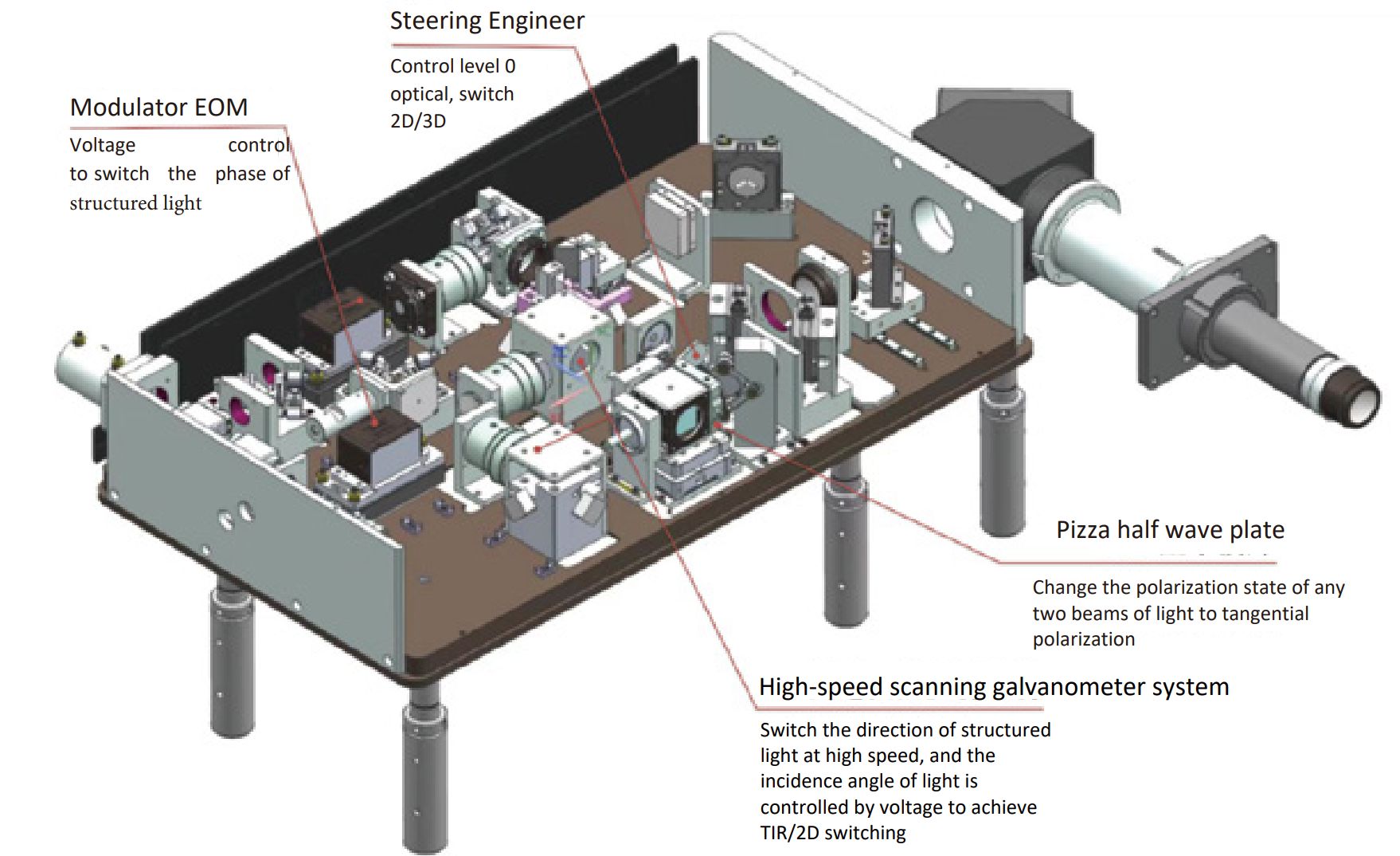
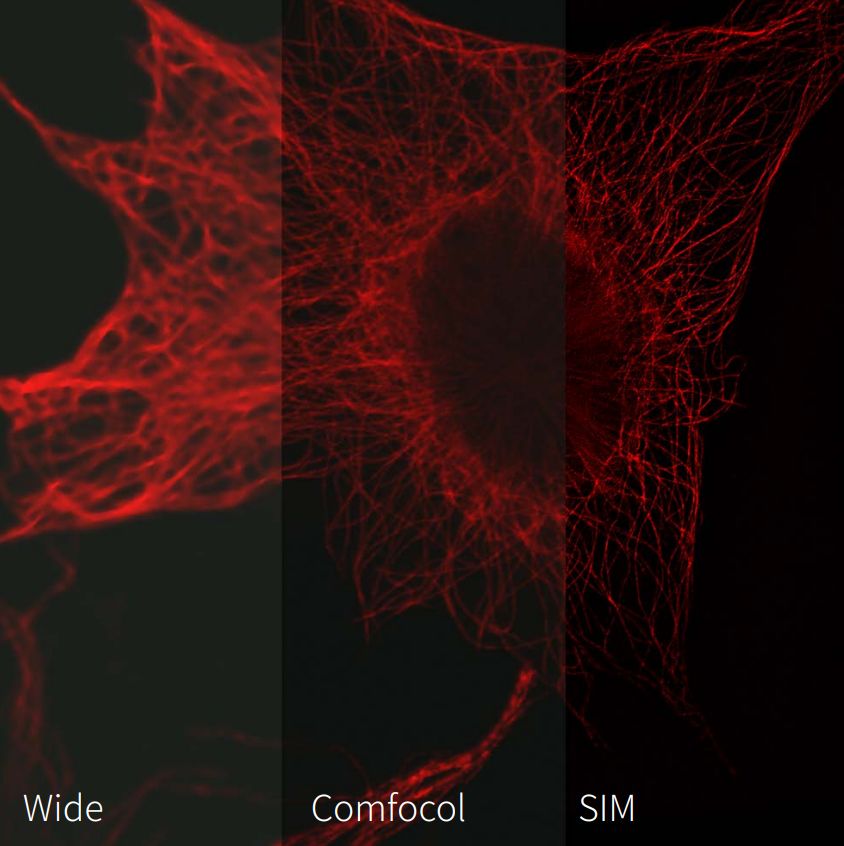
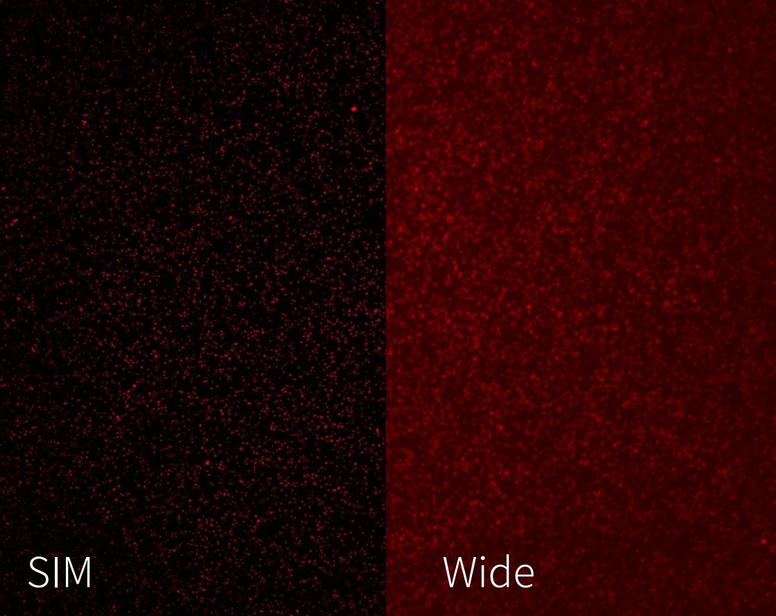
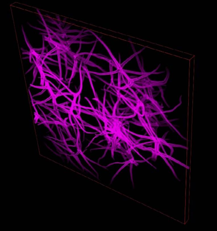
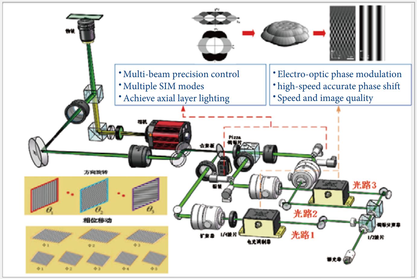
| SIM Illuminator | SIM special structural illumination;achieves twice the optical resolution of traditional microscopy:86 nm in XY,269 nm in Z;real-time super-resolution 25 fps;acquisition speed up to 120 fps |
| SIM Filter Wheel | Electric filter wheel |
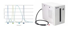
| SIM Dichroic Mirrors | Dichroic mirror 488 nm |
| Dichroic mirror 561 nm |
| Dichroic mirror 640 nm |
| Camera(SIM) | sCMOS camera,NEXCAM-SCMOS 5 |
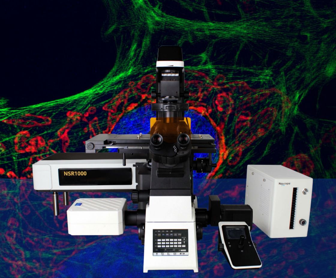
| Main Microscope | NIB1000-AT full motorized inverted microscope |
| Optical System | NIS60 infinite optical system(F200) |
| Eyepiece | 10×,adjustable diopter−5 to+5 |
| Viewing Tube | Tilting 10°–40°,interpupillary distance 47–78 mm |
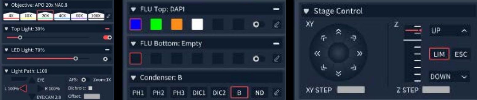
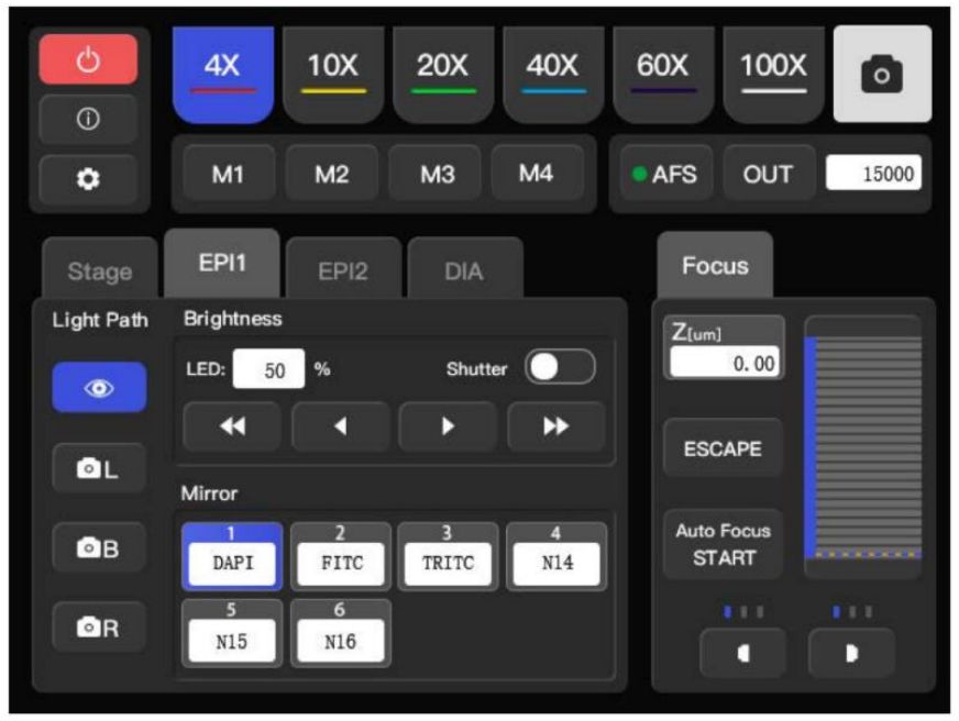
| Objectives(NIS60) | 10×APO,NA 0.45,WD 4 mm |
| 20×APO,NA 0.75,WD 1.1 mm |
| 40×APO,NA 0.95,WD 0.19–0.21 mm |
| 60×APO,NA 1.42,WD 0.25 mm |
| 100×APO,NA 1.45,WD 0.13 mm(Optional) |
| 100×APO,NA 1.49,WD 0.09–0.16 mm(Optional) |
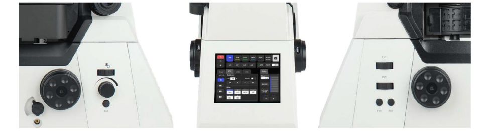
| Nosepiece | Motorized sextuple nosepiece |
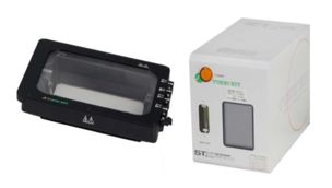
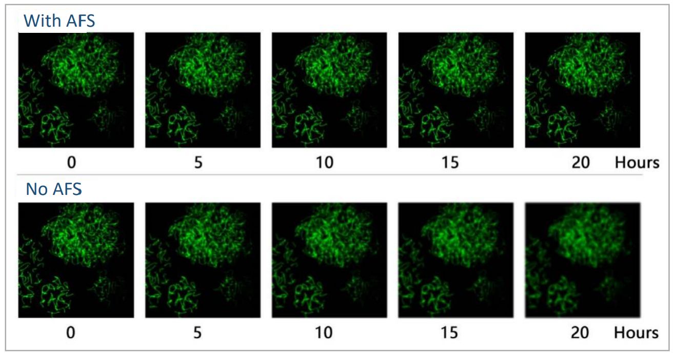
| Motorized sextuple nosepiece with AFS(Optional) |
| Condenser | 7-position motorized condenser |
| Illumination(Transmission) | Köhler illumination,LED |
| Illumination(Fluorescence) | Wide-field 4-channel LED illumination;6-position motorized fluorescence turret(DAPI,FITC,TRITC filter as standard) |
| Intermediate Magnification | Manual 1×,1.5× |
| Output Port | Splitting ratio:Eye 100%,Left port 100%,Right port 100%;Eye 20%/Right port 80% |
| Motorized Stage | Travel range 130×100 mm(stage size 445×300 mm);max speed 25 mm/s;resolution 0.1μm;repeatability 0.5μm |
| Stage Accessories | Universal holder for 35–65 mm culture dishes and slides;optional DIC plate |
| Focusing System | Motorized Z-axis;travel range 10 mm;min step 0.02μm;repeatability 0.1μm;3-step focus knob(2/40/200μm per turn) |
| DIC Plates | 10×,20×,40×,60×,100×plates for nosepiece DIC slot(Optional) |
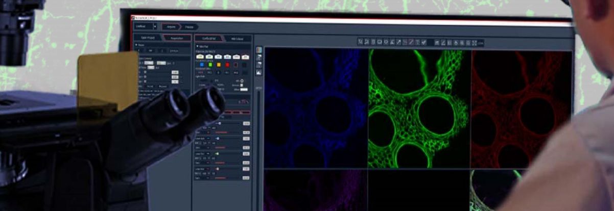
| Software | Nomis Pro X-C |
| Software Functions | Confocal scanning,multidimensional imaging,sub-channel presets,wide-field and confocal imaging,full hardware control,basic image filtering,multiple image output formats |
| Camera(Widefield) | NEXCAM-T12 |
| Extended Functions | Live cell culture system;double-layer fluorescence accessories |