


Phage display is an important laboratory technique for the study of protein-protein, protein-peptide, and protein-DNA interactions that uses bacteriophages (viruses that infect bacteria) to insert proteins of interest within the genetic information that encodes them, displaying the target protein in vitro.
Since its creation, phage display technology has become an important research tool in biological research, fundamentally changing the traditional monoclonal antibody preparation process (hybridoma technology), and is widely used in antigen-antibody library establishment, drug design, vaccine research, and pathogen detection. With the continuous development and improvement of the technology, it has also evolved into a variety of display techniques, such as ribosome display, mRNA display, bacterial display and yeast display, etc.
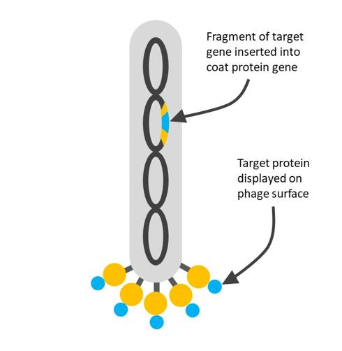 Fig.1. The schematic diagram of phage display technology.
In this technique, a gene encoding a protein of interest is inserted into a phage coat protein gene, causing the phage to "display" the protein on its outside while containing the gene for the protein on its inside, resulting in a connection between genotype and phenotype. These displaying phages can then be screened against other proteins, peptides, or DNA sequences, in order to detect the interaction between the displayed protein and those other molecules. In this way, large libraries of proteins can be screened and amplified in a process called in vitro selection, which is analogous to natural selection.
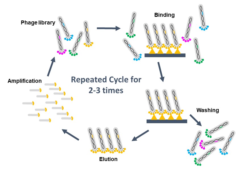 Fig.2 The whole process of phage display technology.
-
Construction of phage display library
Aimed genes are inserted into the multiple phages by recombinant DNA technology so that the protein will be displayed outside of phage particles. -
Binding
Immobilize the target proteins in the wells of the microtiter plate, and add the phage display library to it. In the process, the phage-displaying protein which can interact with the target protein bind on the plate. -
Washing and elution
Phages unbounded to the target proteins can be washed away, and only those showing an affinity for the receptors are left. The left phages attached to the dish can be eluted and used to create more phages by infection of suitable bacterial hosts. -
Amplification
Infect new host cells with those eluted phages to amplify the amount. Repeat the steps above (binding, washing, and elution) several times, further enriching the phage library in binding proteins. -
DNA sequencing
The DNA within the interacting phage is sequenced to identify the interacting proteins or protein fragments.
- Large functional size with high diversity
- Allowing the automation of panning processes and the amplification of expression levels
- Convenient to cooperate with any species
- Analysis of protein binding properties
- Monoclonal antibody specificity profiling
- Analysis of protein post-translational modifications
- Establishment of antigen-antibody library
- Pathogen detection
- Gene therapy
- Research on host-microbe interactions
- Biomarker identification
- Cell signaling research
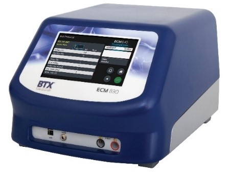
Electroporators
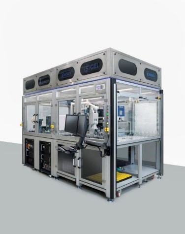
Lab Automated Extraction Workcells
Pipettes
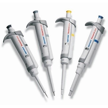
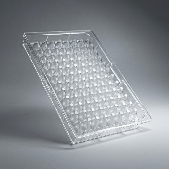
Microtiter Plates
Microplate Washers
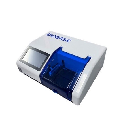
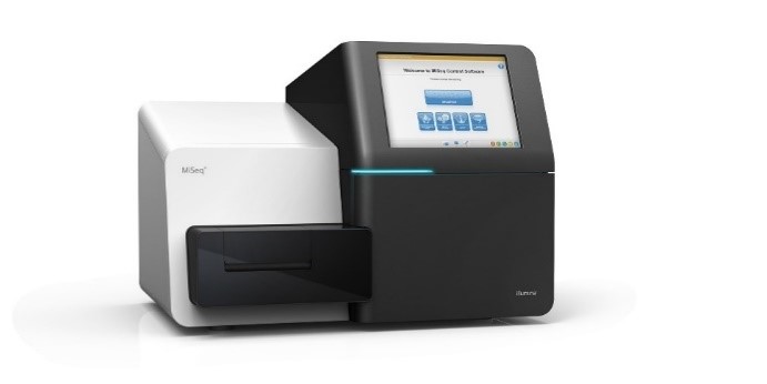
DNA Sequencing Equipment
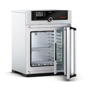
Laboratory Incubators
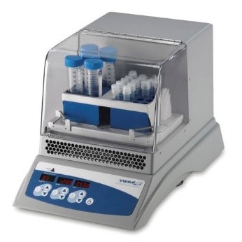
Laboratory Rockers and Shakers
Centrifuges
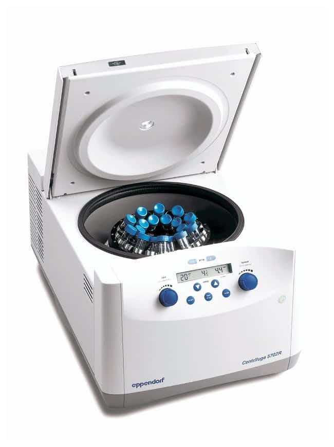
STEMart provides you with a variety of equipment or consumables used in phage display technology to meet your various R&D and application needs. If you have any questions or requirements for phage display technology, please feel free to contact us.