SUPER VISION
Obtain large viewing field and whole sample information in a short time
There is no product in the shopping cart.
Super vision, super resolution, and super speed
As life science research continues to expand toward more complex tissues,organs,and biological models,advanced imaging performance has become essential for scientific discovery.Studying large and intricate samples requires both a wider imaging area and faster acquisition to capture subtle intracellular activity with high fidelity.The NCF1000 confocal laser microscope was developed to address these challenges by delivering high-throughput,high-quality imaging for modern research environments.
Featuring a class-leading 25 mm field of view,the NCF1000 enables seamless imaging of large specimens in a single scan.With a maximum scan resolution of 8192×8192 pixels,it produces sharp and detailed images even when using low-magnification objectives.At the same time,imaging speeds of up to 60 frames per second(8×256 pixels)allow researchers to combine wide-area coverage with high-resolution capture,generating more data in less time.
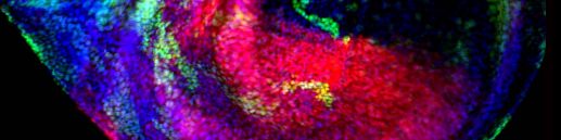
By integrating a wider field of view,higher spatial resolution,and faster acquisition,the NCF1000 delivers clearer images,enhanced contrast,and more accurate color representation.This combination allows researchers to explore cellular structures at the nanoscale,observe biological processes with greater precision,and push the boundaries of life science research into previously unexplored territory.
Obtain large viewing field and whole sample information in a short time
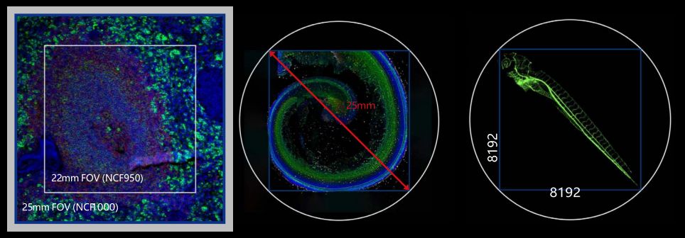
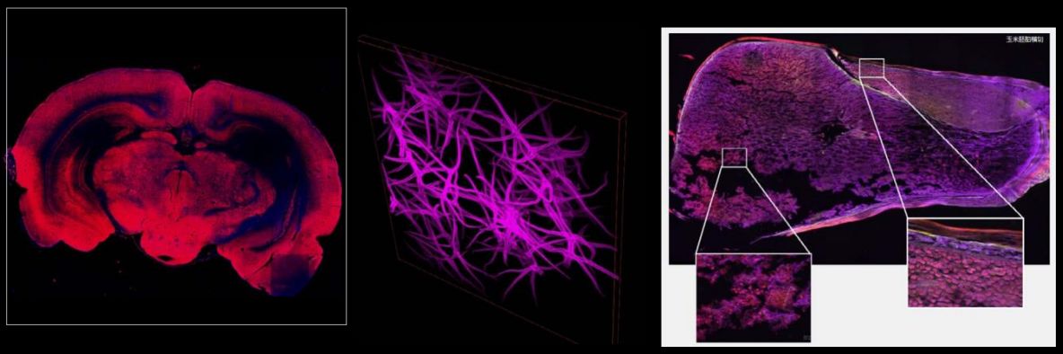
With its advanced 25 mm field of view,the NCF1000 confocal microscope can image large specimens in a single acquisition,delivering up to 1.5×higher data throughput compared with earlier microscope generations.When combined with scanning resolutions of up to 8192×8192 pixels,the system meets the growing need for detailed analysis of tissues,organs,and living or model organisms,providing rich biological data for life science research.
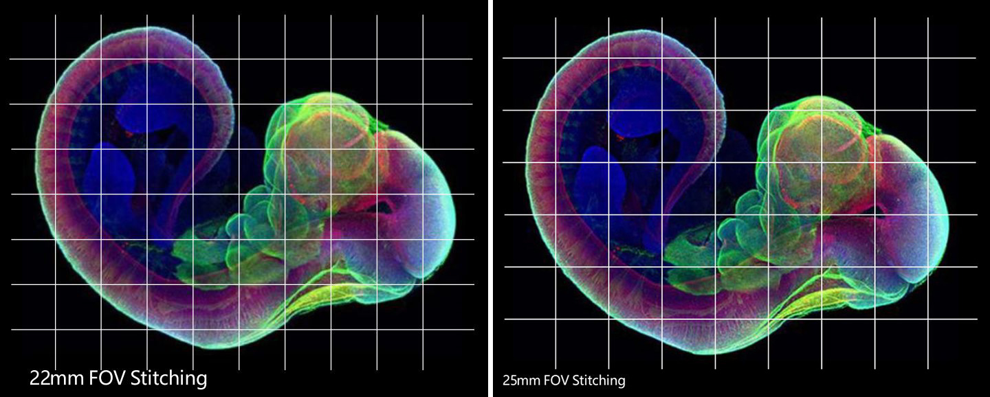
When paired with the NIB1000 inverted microscope,the NCF1000 can generate high-quality confocal images across a full 25 mm field of view.The expanded imaging area significantly reduces the number of individual frames needed for image stitching,shortening acquisition time while maintaining uniform brightness,high efficiency,and high-throughput performance for large samples.
Obtain higher SNR and higher image resolution
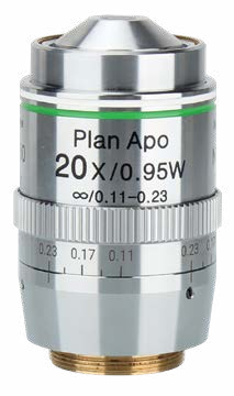
NIS Series objectives are engineered with high numerical aperture,extended working distance,and advanced chromatic aberration correction.Their multi-layer optical coatings deliver superior image clarity and resolution.In addition to serving as an excellent choice for conventional optical microscopes,NIS objectives are also a critical component in achieving high-performance confocal imaging.

NIS Apochromat TIRF 100×Oil(100×APOTIRF)
With an ultra-high numerical aperture of 1.49,this objective provides outstanding resolution and represents the highest level of the NIS series.It is designed to correct spherical aberrations at both 23°C and 37°C for stable imaging under different temperature conditions.
NIS Apochromat 20×CWI(20×APO Water)
Designed for observing cultured cells and specimens in aqueous media,this objective matches refractive indices close to those of cells and culture solutions,helping to reduce spherical aberration and light loss caused by refractive index mismatch.
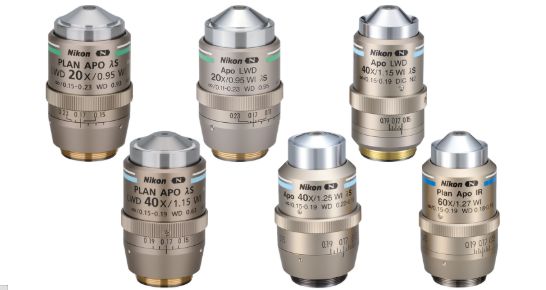
NIS Plan Apochromat Series(APO)
This professional-grade objective line pushes the limits of numerical aperture and working distance,delivering precise aberration correction across the full field of view and image quality beyond what conventional objectives can achieve.
NIS Series objectives are designed with high numerical aperture,long working distance,and advanced chromatic aberration correction.Their multi-layer optical coatings deliver high image clarity and resolution,making them well suited for both standard optical microscopes and confocal imaging platforms.
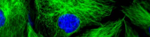
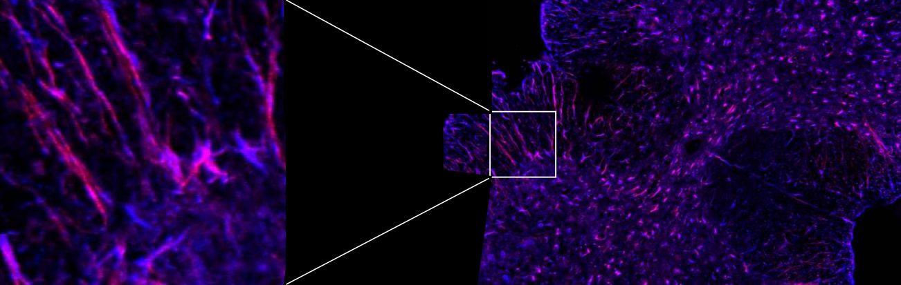
The NCF1000 integrates a high-precision galvanometer scanning system and a continuously adjustable electronic pinhole within the scan head to achieve low noise,high contrast,and high-quality confocal imaging.With a maximum scan size of 8192×8192 pixels,even low-magnification objectives can provide fine spatial sampling,ensuring accurate capture and faithful reproduction of microscopic details.
Acquisition time reduction High frequency confocal imaging is realized
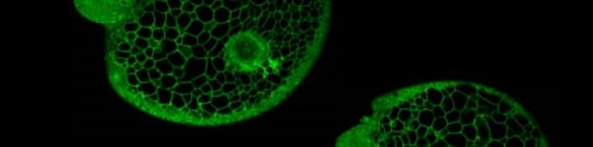
Real-time four-channel synchronous imaging with frame rates up to 60 fps(8×256 pixels)shortens exposure time under high illumination and greatly reduces phototoxic effects.The high-speed acquisition enables continuous,high-frequency data capture to record dynamic events and long-term sample changes in real time,supporting the demanding imaging requirements of life science research.
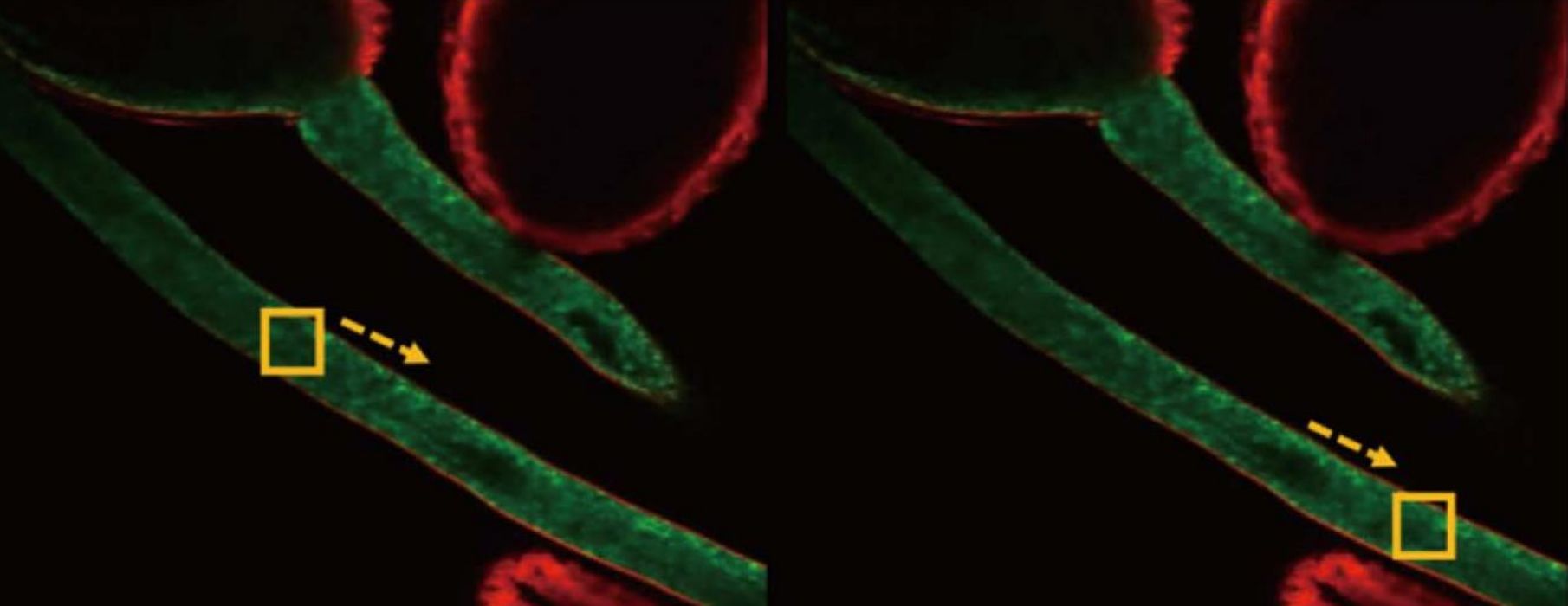
With imaging speeds of up to 60 fps(8×256 pixels),the NCF1000 minimizes exposure under high illumination and significantly lowers phototoxic effects.Its high-speed acquisition supports continuous data collection,enabling accurate recording of dynamic processes and long-term changes to meet the demanding needs of life science imaging.
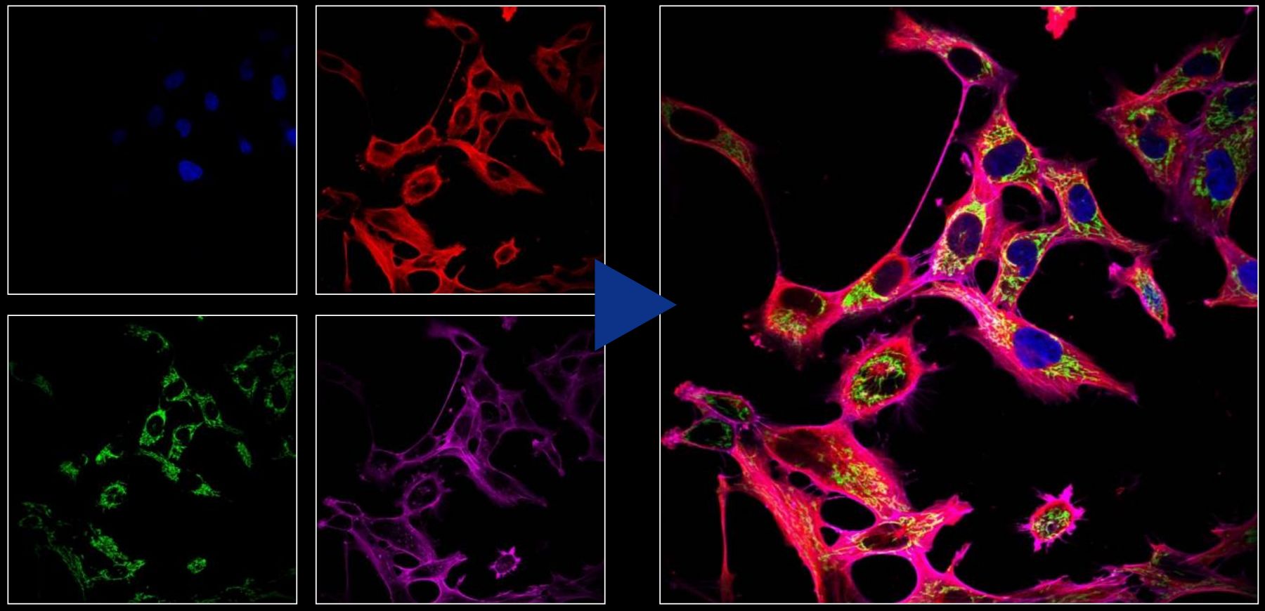
The NCF1000 features advanced four-channel fluorescence fusion technology,allowing multiple fluorescence signals to be captured and combined simultaneously in real time.Four different fluorescent labels can be detected and analyzed within the same field of view,with seamless channel switching and spectral separation,revealing complex multi-dimensional information while improving experimental efficiency and data accuracy.
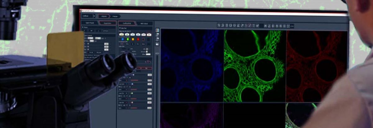
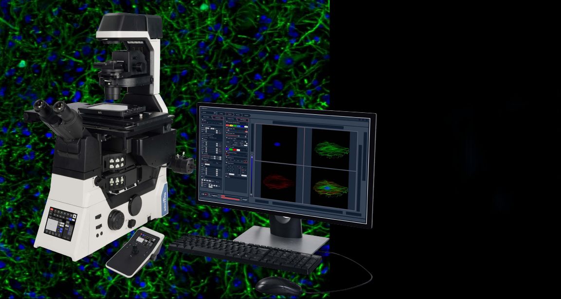
Powerful software for analysis
The system provides intuitive digital management and precise control of multiple motorized components within the microscope,including objective switching,focusing,condenser selection,and fluorescence module changes.
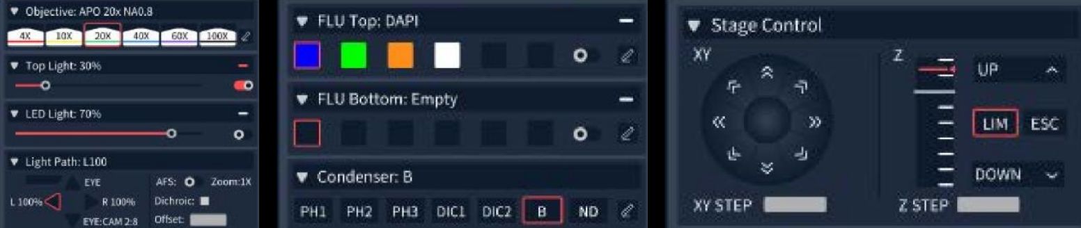
User-defined observation modes can be stored,supporting combined scanning across X,Y,Z,λ,and T dimensions.A range of flexible acquisition modes is built in,including multi-channel fluorescence imaging,time-lapse recording,multi-position capture,Z-stack imaging,and panoramic stitching.These modes can be freely combined to suit a wide variety of complex experimental applications.
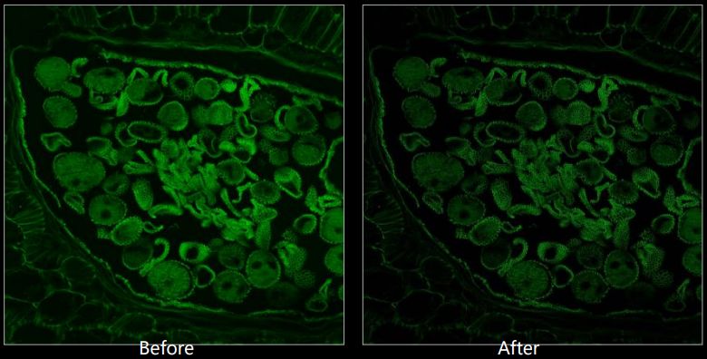
Two-dimensional images can be sharpened through deconvolution,with repeated processing available to reduce shot noise in confocal data.Three-dimensional deconvolution is also supported for multidimensional image sets.
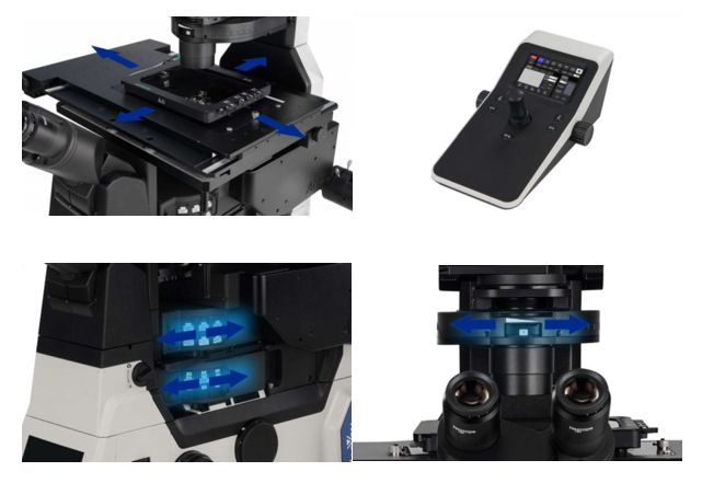
The NIB1000 serves as a versatile and high-powered imaging platform for the NCF1000 system,providing a stable and expandable solution for advanced microscopy.Its 25 mm field of view supports efficient imaging of large specimens and enables high-throughput workflows.
Multiple imaging modes—including brightfield,fluorescence,DIC,and phase contrast—are integrated into the system.Users can choose between single-or dual-layer optical paths to optimize performance for a wide range of experimental requirements.
Designed for live cell observation,the system precisely controls the temperature,humidity,and CO₂levels of the sample stage,creating a stable and suitable environment for long-term experiments.
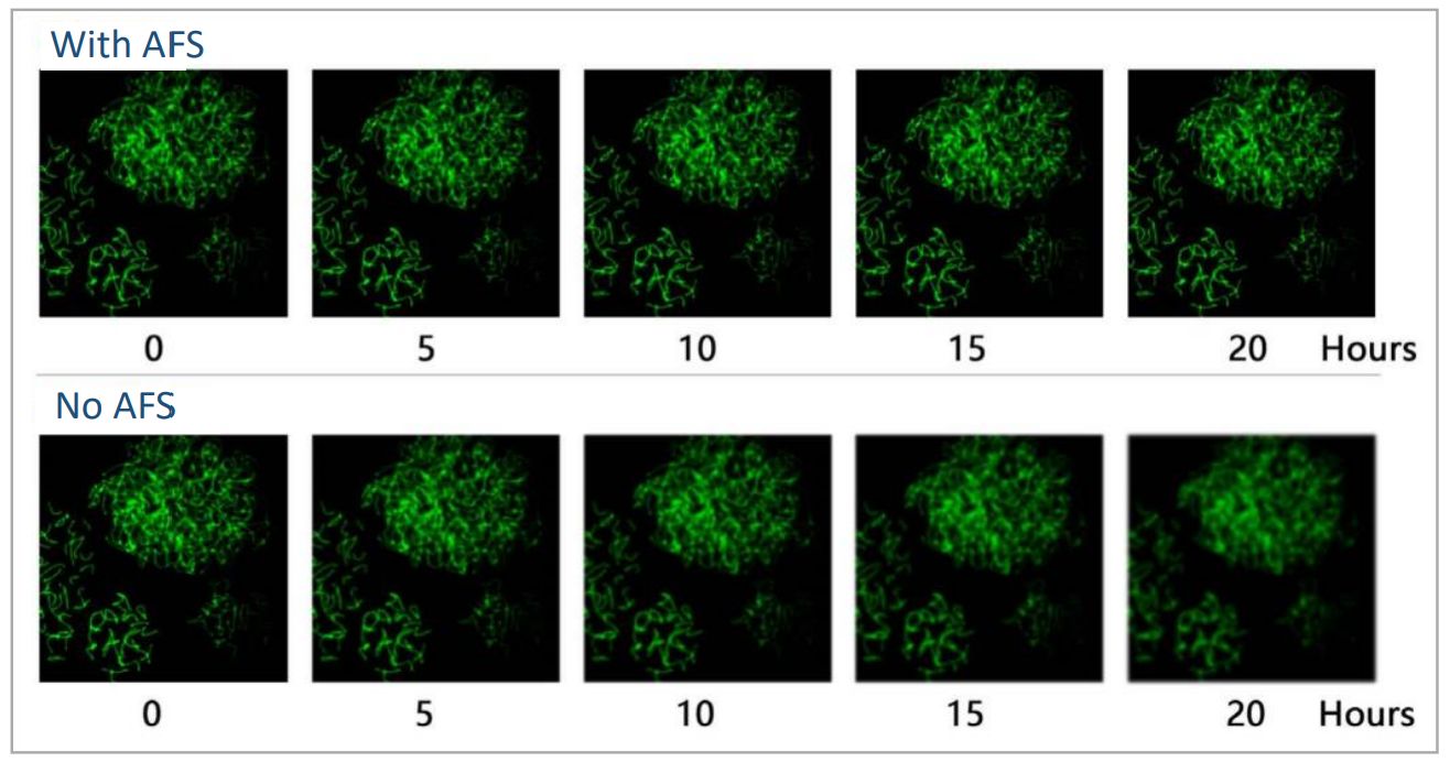
The NIB1000 features an independent focusing mechanism that minimizes the influence of other mechanical components on the Z-axis.Its Adaptive Focus System automatically corrects focus drift,ensuring sharp and stable images whether using high-NA objectives or advanced techniques such as super-resolution,confocal,or TIRF imaging.
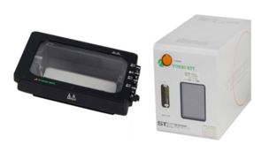
The LED4 light source provides up to four independent illumination channels with high excitation efficiency and strong brightness to support routine fluorescence imaging.It offers long service life with continuous operation,eliminates the need for bulb replacement,and helps reduce photobleaching and phototoxicity for sensitive samples.
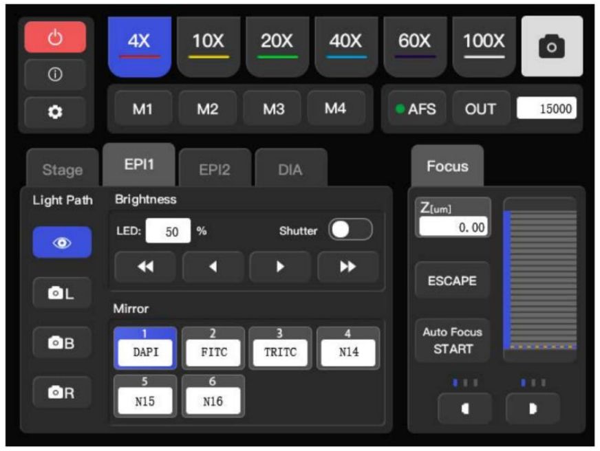
The front panel of the NIB1000 incorporates a touch screen that enhances usability and system expandability,while traditional control knobs and buttons are retained on both sides for convenient operation.This design allows easy control even in low-light environments,enabling researchers to focus on their experiments without complex adjustments.
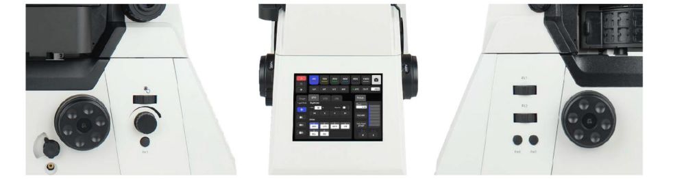
The 5.6-inch touch display allows direct control of key motorized components,including objective switching,fluorescence filter selection,condenser light intensity,stage and Z-axis movement,beam splitter settings,and user-defined function keys.It also shows real-time information such as objective magnification,illumination level,active fluorescence channel,output port,and XYZ position and speed.
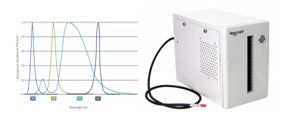
The system uses a large-aperture tube lens to significantly increase light throughput,which,when combined with a wide-field CMOS sensor,supports brightfield and fluorescence imaging across a full 25 mm field of view.The fluorescence illumination module is optimized for FOV 25 mm and employs high-power LEDs to deliver a broad,high-transmission excitation spectrum,including ultraviolet wavelengths.Matched large-diameter fluorescence filters ensure high signal-to-noise imaging across the entire field.
| NCF1000 Laser Confocal Microscope Specifications | |
|---|---|
| Item | Specification |
| Laser Unit | 405 nm,488 nm,561 nm,640 nm |
| Standard Detector | 400–750 nm,4 PMT |
| Transmission Detector | Single-channel PMT detector |
| Scanner | Max pixel:8192×8192 |
| Scan Modes | X,Y,Z,λ,T,and multi-mode combinations |
| Pinhole | Stepless adjustment |
| Confocal Field of View | Φ25 mm enclosed square |
| Image Bit Depth | 16 bits |
| Motorized Microscope | NIB1000-AT motorized research microscope |
| Optical System | NIS60 infinite optical system |
| Eyepiece | 10×,diopter adjustment−5 to+5 |
| Viewing Head | Tilting 10°–40°,interpupillary distance 47–78 mm |
| Objectives(NIS60) | 10×APO,NA 0.45,WD 4 mm |
| 20×APO,NA 0.75,WD 1.1 mm | |
| 40×APO,NA 0.95,WD 0.19–0.21 mm | |
| 60×APO,NA 1.42,WD 0.25 mm | |
| 100×APO,NA 1.45,WD 0.13 mm(Optional) | |
| 100×APO,NA 1.49,WD 0.09–0.16 mm(Optional) | |
| Nosepiece | Motorized sextuple nosepiece with DIC slot |
| Motorized sextuple nosepiece with DIC slot and AFS focus maintain unit(Optional) | |
| Condenser | 7-position motorized condenser |
| Transmission Illumination | Köhler illumination,LED |
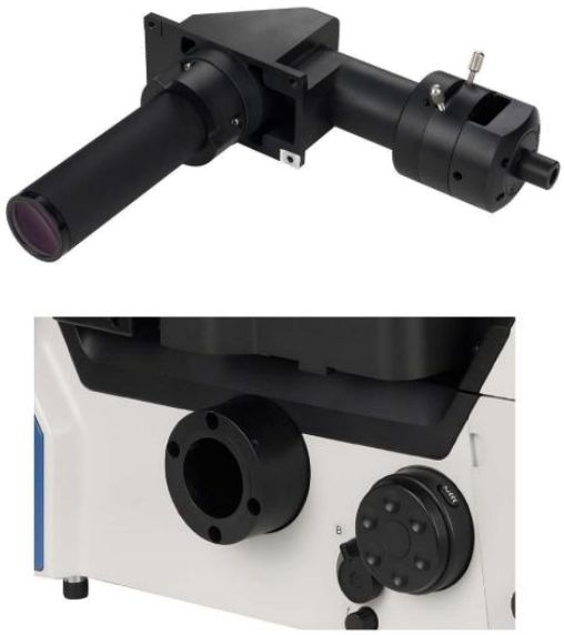
| EPI-Illumination | 4-channel LED illumination;6-position fluorescence turret(standard BGU filter cube);motorized shutter |
| Intermediate Magnification | 1×,1.5× |
| Beam Splitter | Eyepiece 100%;Left port 100%;Right port 100%;Eyepiece 20%/Right port 80% |
| Motorized Stage | Travel range 130×100 mm(stage size 445×300 mm);max speed 25 mm/s;resolution 0.1μm;repeatability 0.5μm |
| Stage Accessories | Universal holder for 35–65 mm Petri dishes and slides;well clamp holder |
| Motorized Focusing System | Motorized Z-axis;travel range 10 mm;min step 0.02μm;repeatability 0.1μm |
| Focus Knob | 3-step focus knob:2μm/turn,40μm/turn,200μm/turn |
| DIC Plates | 10×,20×,40×,60×,100×DIC plates for nosepiece DIC slot(Optional) |
| Software | Nomis Pro X-C |
| Software Functions | Confocal scanning,multidimensional imaging,sub-channel presets,widefield and confocal imaging,full hardware control,basic image filtering,multiple image output formats |
| Camera | Nexcam-T12 |
| Optional Systems | Live cell culture system;SR-SIM super-resolution system;2-layer optical system |
To learn how the NCF1000 Confocal Microscope can be configured for your specific imaging workflow,contact usto discuss your samples,imaging requirements,and system options.
Our application specialists will help you select the optimal configuration,accessories,and software to support your research with maximum performance and efficiency.